Cochlea of the Inner Ear: A Model of Its Dynamics
by J. J. Zwislocki
Computer Program by E. J. Kletsky and D. J. Arpajian
Reference. Jozef J. Zwislocki, Auditory Sound Transmission, Lawrence Erlbaum Associates, Mahwah, New Jersey.
The cochlea is the auditory end organ located in the inner ear. It consists of a snail-like, spiral canal in the temporal bone of the skull. It is filled with fluid that is practically incompressible and is divided longitudinally in two by a visco - elastic partition. Transversal waves are generated on the partition by sound when it enters the canal at its basal end through a small opening, called the oval window. The associated fluid pressure is equalized on the opposite side of the partition through another opening, called the round window. The partition is interrupted by a small opening, the helicotrema, at the other end of the cochlea, called the apex. Only at very low sound frequencies do the cochlear waves reach the apex. As shown in the cross-sectional sketch of the canal, the partition comprises 4 mechanically significant structures:
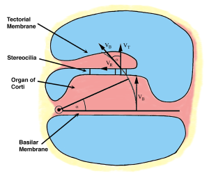
- An elastic plate called the basilar membrane
- A bulky cell mass, called the organ of Corti, that contains 4 longitudinal rows of the auditory sensory cells — 3 rows of outer hair cells and one row of inner hair cells
- Bundles of stiff stereocilia, popularly called hairs, projecting from the hair cells. They are indicated by thick vertical lines protruding from the upper surface of the organ of Corti, called the reticular lamina
- An acellular mass, the tectorial membrane, suspended viscoelastically from the bone surrounding the cochlea. It is coupled viscoelastically to the organ of Corti by the stereocilia bundles of the outer hair cells. The stereocilia of the inner hair cells have been found to play a negligible mechanical role.
Sound waves produce an up-and-down oscillation of the partition according to a characteristic pattern dictated mainly by the stiffness properties of the basilar membrane and the cellular structures within the organ of Corti. The basilar membrane motion approaches in cross section that of a stiff beam rotating around its inner edge. The rotation generates a tangential component at the reticular lamina, designated as “radial”, which entrains the relatively soft tectorial membrane through the streocilia bundles. The force required to move the tectorial membrane deflects the stereocilia bundles radially. Depending on the direction, the deflection produces either depolarization or hyperpolarization of the hair cells and stimulates the fiber endings of the auditory nerve. The alternating polarization produces an alternating electric field at the hair cells. The outer hair cells respond to this field by alternating axial contractions and dilations that are converted to alternating stereocilia deflections. In this way, a positive feedback loop is created in the outer hair cells that act as both mechano-electric and electromechanical transducers. The feedback enhances the radial vibration of the tectorial membrane and, through it, the transversal vibration of the basilar membrane.
The cochlear partition can be modeled in its cross section as a system of two coupled resonators, one consisting of the cell mass supported by the viscoelastic basilar membrane, the other, of the mass of the tectorial membrane also supported viscoelastically. Both supports are anchored to the bony wall of the cochlea. The two resonators are coupled transversally through the viscoelastic stereocilia and longitudinally through the cochlear fluid. A system of two coupled resonators has two resonance frequencies. In the cochlea, these frequencies are relatively low at the apex, and increase toward the base, mainly because of gradual stiffening of the viscoelastic elements. The second maximum is severely decreased by viscosity.
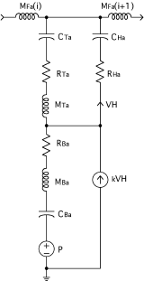
The theory of electroacoustic analogies enables us to express the cochlear system described above in terms of an electrical network. In the network, the voltage source, P, models the sound-pressure difference across the cochlear partition; the capacitance, CBa, and the resistance, RBa, the compliance and viscous resistance of the basilar membrane, respectively; the inductance, MBa, the effective cell mass resting on the basilar membrane; MTa, the effective tectorial-membrane mass; CTa and RTa, the viscoelastic support of this mass; CHa and RHa, the viscoelastic parameters of the stereocilia bundles; VH, the volume velocity of the stereocilia deflection; kVH, the feedback volume velocity generated by the electro-motility of the outer hair cells. The two inductances, MFa, model the longitudinal fluid coupling.
In the computer simulation, the cochlea is modeled by a transmission line consisting of a ladder network of these resonators coupled through the inductances MFa(i).